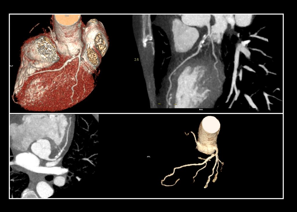
FUH Medical Review
Share this article:

Undergoing a complex surgery, that too of the heart, can be a scary experience for patients. On most occasions, patients try and find ways to avoid a surgery episode. Well, the good news is, in some cardiac patients, surgery avoidance is now possible.

The case

This is about a 52-year-old female patient, who experienced chest pain and shortness of breath, after the second dose of COVID-19 vaccine. She had a past medical history of dyslipidemia, depression, and anorexia nervosa. Upon initial examination, ECG delineate SR and Poor R wave progression in anterior leads and ST&T inversion in inferolateral leads were seen. ECHO was showing good LVSF. Her temperature was 36.3C, pulse 102/beats/mins and BP was 102/61.

Based on her clinical presentation and cardiovascular risk factors, she was referred to Dr. Syed Sakib, Specialist Interventional Cardiologist at Fakeeh University Hospital, for coronary computed tomography angiography (CTCA) and further evaluation.

Generally, arrhythmia (which is any disturbance of the normal rhythmic beating of the heart or myocardial contraction), is a hurdle to acquire best images in a cardiac imaging process. However, advancement in CT technologies at Fakeeh University Hospital help the clinical team to overcome such challenges. In this case, advance CT scanner technical capabilities such as Dual Source Acquisition and Native Temporal resolution, helped the team to overcome such high heart rate CT imaging challenges.

Course of Intervention

Dr. Sakib and his team, Masood Ahmed-Radiology Manager and Dr. Latha PK-Specialist Radiologist, performed a study with cardiac ultra-fast imaging (Flash technology) to minimize the motion artifact and reduce the dose to as low as possible. Study was performed with low kv flash calcium scoring and contrast timing was calculated with test bolus technique. Coronary CTA was performed with high pitch flash technique with 50ml of contrast and 40ml of saline at a flow rate of 5.5ml/sec.

Results Seen by the Team

Dominance

Left dominance supplied by branches of LAD, LCX. Left Main divides into LAD, Ramus, LCX and LAD. Curved MPR images depict irregular mixed density plaque with calcification in proximal segment of LAD, causing luminal moderate stenosis. A total of three calcific plaques are delineated with CTCA with normal opacification in distal to it. Diagonal and RAMUS appears to be normal. Left circumflex depict diffuse mild narrowed caliber. Obtuse marginals OM1 and OM2 are patent. Right coronary artery shows diffuse moderate to severely narrowed caliber of proximal segment, severely narrowed in mid segment.

Conclusion

Variability in heart rhythm (bradycardia, tachycardia, arrythmia etc.) in the coronary arteries, make diagnostic evaluation very unpredictable and mostly unreliable with conventional technologies. Use of advanced dual source CT technologies (with AI algorithms for heartrate variability, fastest rotation time, high native temporal resolution, high pitch low dose acquisition in conjunction with ECG-gating), to reconstruct images in the optimal phase of the cardiac cycle, are essential for sharp depiction of the coronary arteries with minimum blurring from the cardiac motion.

This requirement is particularly critical in patients with high and variable heart rates. The technology improves the diagnostic quality for coronary artery diseases of such patients. In addition to diagnosing these challenging clinical conditions, Dual Source technology helps to scan such patients with an ultra-low dose.

Dr. Syed Sakib

Specialist Interventional Cardiologist
Fakeeh University Hospital, Dubai

Dr. Syed Sakib

Specialist Interventional Cardiologist
Fakeeh University Hospital, Dubai

By using the right kind of advanced technology for the patient, many doctors and hospitals can save the patient from a possible surgery.

Share this article:

Contribute

For the next FUH Medical Review

Subscribe

To FUH Medical Review